


Sydney Podiatric Surgery – Specialist Podiatrist
What Is an Accessory Navicular?
An accessory navicular is an extra piece of bone located on the inside of the foot, just above the arch. It is also known as an os naviculare or os tibiale externum. This is a common anatomical variation, occurring in approximately 10–12% of the population, and many people never realise they have one.
During childhood, the accessory navicular begins as cartilage and gradually hardens into bone as the skeleton matures. In some people, this creates a noticeable bony prominence on the inside of the foot. While it is often painless, it can become irritated by footwear, prolonged walking, running, or sporting activities, leading to discomfort and inflammation.
The accessory navicular is closely connected to the tibialis posterior tendon, one of the most important structures supporting the arch of the foot. Because of this attachment, irritation of the accessory navicular can also affect the function of the tendon, making activities such as walking, running, or standing for long periods more uncomfortable.
An accessory navicular can be found in people with both normal foot posture and flat feet (pes planus). Those with flatter feet may place greater stress on the tendon and accessory bone, increasing the likelihood of symptoms.
Types of Accessory Navicular
Accessory navicular bones are classified into three main types based on their size, shape, and attachment to the main navicular bone.
- Type I: A small or oval bone embedded within the tibialis posterior tendon.
- Type II: The most common symptomatic type. It is typically larger than type I and it is connected to the navicular bone by a layer of cartilage or fibrous tissue (rather than solid bone). Irritation of this fibrous attachment through movement can typically cause pain.
- Type III: Occurs when a Type II accessory navicular fuses completely with the main navicular bone. This creates a large bony prominence on the inside of the arch which can cause pain via rubbing and irritation on shoes and activities.
Surgical Options for Accessory Navicular in Sydney
Removal of the accessory navicular is a procedure that is commonly referred to as a Kidner Procedure, however there are some variations to this procedure.
In some cases where the accessory bone is type I or II (separate from the main navicular body) it can be easily removed with very minimal detachment of tibialis posterior tendon. This can be performed in many cases, patients are placed in a CAM boot and will not weight bear for 2–3 weeks. Partial weight bearing is then commenced and the patient is transitioned into a good stable shoe. Postoperative rehabilitation is then commenced to help strengthen the arch and tendons that support the arch.
In some cases in which the accessory bone is large or the patient has symptoms associated with a flatfoot or tendon weakness, then once the accessory bone is removed the tendon is reattached to the main navicular bone helping to strengthen the arch. This may require the use of an anchor to provide further stability and strength. In such cases, the patient will require a longer recovery as the foot will be immobilised to allow for adequate healing of the tendon given it was reattached.
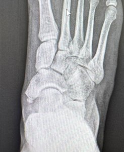
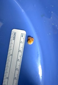
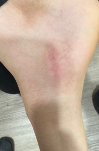
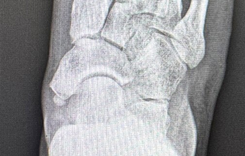
Figure 1: Pre op x-rays showing the accessory navicular bone
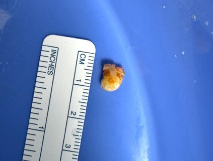
Figure 2: Intra operative picture showing the excision of the accessory bone
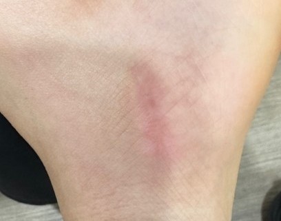
Figure 3: Post-operative picture of incision location/scar healing
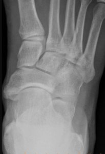
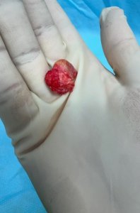
Figure 4: A pre operative x ray showing a larger type III (cornuate) accessory navicular
Figure 5: The accessory bone excised
Do you want to know more about this procedure?