


Surgical treatment for painful posterior heel spurs and Achilles tendon problems
Pain at the back of the heel can be particularly frustrating, especially when it interferes with walking, running, exercise or wearing normal shoes. In some patients, the pain is caused by a combination of a prominent heel bone and degeneration of the Achilles tendon where it attaches to the heel. Some common pathologies that cause this include a Haglund’s deformity and/or retrocalcaneal (back of heel bone) spurring. When conservative treatment has failed, surgery may be indicated. Surgery generally involves removing the bone prominence and repairing the damaged tendon.
What Is a Haglund’s Deformity?
A Haglund’s deformity is a prominent area of bone at the back of the heel bone (calcaneus). This prominence can irritate the surrounding soft tissues and Achilles tendon, particularly when wearing shoes with a firm heel counter. Over time, repeated irritation can contribute to inflammation and degeneration of the Achilles tendon.
What Is a Retrocalcaneal Exostosis or Heel Spur?
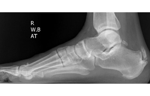
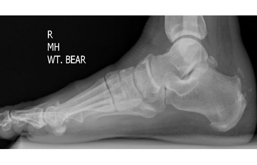
A retrocalcaneal exostosis is a growth of bone at the back of the heel bone. This spur or growth of bone can increase in size with time and can irritate the surrounding structures. In some patients the spur can grow within the Achilles tendon, causing pain and degeneration of the Achilles tendon. The bony prominence can become large enough that it causes irritation due to excessive rubbing on footwear.
Patients may experience:
- Pain at the back of the heel
- Swelling around the Achilles tendon
- A noticeable bony prominence
- Pain when wearing certain shoes
- Stiffness in the Achilles tendon
- Pain during walking or running
- Difficulty performing activities such as running or jumping
- Tenderness where the Achilles tendon attaches to the heel
Conservative Treatment for Posterior Heel Pain
Surgery is generally considered only after appropriate non-surgical treatment has been attempted. Conservative care for posterior heel pain can include activity modification, footwear modifications, custom orthoses, heel lifts, Achilles tendon strengthening and stretching, and injection therapy (PRP, corticosteroid).
Surgery is more likely to be considered when there is:
- A significant posterior heel prominence
- Persistent insertional Achilles pain
- Significant Achilles tendon degeneration
- Bone spurs at the Achilles insertion
- Difficulty with normal footwear
- Ongoing limitation with exercise or sport
What Is Involved in Retrocalcaneal or Posterior Heel Spur Removal?
Before surgery is considered, imaging such as X-rays, ultrasound and/or MRI may be used to assess the bone and Achilles tendon. During the consultation, the specialist will then decide which procedure is best suited to your condition.
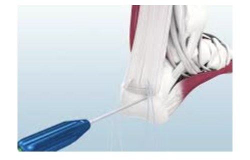
Retrocalcaneal exostectomy or heel spur surgery involves performing an incision on the back of the heel over the site of the heel spur. A small incision is performed through the Achilles tendon and a portion of the tendon is reflected off the heel bone. The goal is to reflect only a minimal portion of the Achilles tendon to maintain strength and integrity. Once this is done, the excess bone or spur is carefully removed and the remaining bone smoothed out. If the Achilles tendon has significant degenerative changes, the unhealthy tendon tissue can also be removed.
In some instances, where a significant portion of the Achilles tendon needs to be reflected from the heel to access and treat the underlying pathology, the healthy tendon may need to be re-attached via specialised bone anchors and strong sutures.
Retrocalcaneal exostectomy or heel spur surgery is usually performed as day surgery under general anaesthesia. A thorough clinical work up is performed to ensure your pre-operative health is suitable for this procedure and to ensure minimal complications post-operatively.
Recovery After Achilles Insertional Surgery
Recovery after Achilles re-attachment is more involved than many other foot procedures because the tendon requires time to heal securely to the bone. Patients will generally require a period of immobilisation and protected weight bearing following surgery.
Typically a patient may be placed in a back slab or cast and required to be non weight bearing for 3 weeks. This is then followed by partial weight bearing in a CAM boot for 3 weeks. At this point, patients are slowly transitioned into supportive footwear with a temporary heel lift to help reduce the tension on the tendon. Weight bearing and rehabilitation are then gradually introduced. The recovery process generally involves:
- Initial protection of the Achilles tendon
- Gradual progression of weight bearing
- Restoration of ankle movement
- Progressive calf and Achilles strengthening
- Gradual return to normal footwear
- Gradual return to exercise and sport
It is important not to progress activity too quickly because the Achilles tendon needs adequate time to heal. Patients are expected to make a full recovery and return to pre-surgical activities. Given it is a more extensive procedure than other foot procedures, specific risks such as infection, DVT, wound breakdown and tendon weakness are all assessed and accounted for.
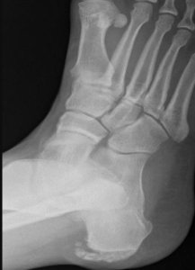
Figures 1 and 2: Pre-operative x-rays showing retrocalcaneal spurring – note that the spur sits within the tendon and has fractured off the body of the calcaneus
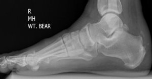
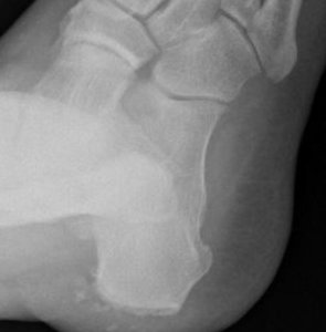
Figures 3 and 4: Post-operative x-rays following resection of the retrocalcaneal spur
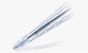
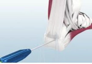
Figures 5 and 6: The Achilles tendon anchored down to the calcaneus using the Y-Knot® PRO RC calcaneus Achilles anchor (supplied by ConMed)
Do you want to know more about this procedure?
Learn more about insertional Achilles tendon pain, what causes it and how it is assessed and treated at Sydney Podiatric Surgery.